
Science with Passion

App-Nr.: VBS0089
Version 1 05/2026
Hey, ho, oliGO – Comparison of ion-pairing systems for oligonucleotide analysis with HPLC-UV
J. Kramer, M. Prüfer, J. Wesolowski, K. Folmert; kramer@knauer.net
KNAUER Wissenschaftliche Geräte GmbH, Hegauer Weg 38, 14163 Berlin
Zusammenfassung
Oligonucleotides (ON) are the new superstars in medical and biological research, especially when it comes to highly specialized or personalized drugs. More drugs are reaching the clinical phase of development or have already been approved by the Food and Drug Administration. The target group is often particularly vulnerable patients, which is why the quality requirements for synthesized oligos and the throughput in analytical laboratories are enormous. The challenge of analysis lies in the very long polar chains, which differ only in individual nucleobases or sometimes in individual functional groups [1].
Einführung
Ion-pairing reversed phase (IP-RP) chromatography is one of the most used methods for the analysis of oligonucleotides (ON). Oligonucleotides are highly customizable compounds. The backbone can be modified to improve identification of each nucleotide to more easily track ON synthesis [2] or to improve protection against degradation by enzymes [3]. The sugar ring can also be modified. Often, modifying the 2’ position on the sugar ring will result in greatly increased binding capabilities while also reducing non-specific protein binding to the oligonucleotide [4]. 2’-O-methyl and 2’-O-methoxyethyl are the most common modifications on the sugar ring. Those short nucleotide sequences are becoming increasingly essential for molecular biology and therapeutic ends. But with their naturally negative charge, it is not that easy to gain retention on a column. That’s where the ion-pairing mechanism comes into play. By adding a positively charged ion pair reagent, the oligonucleotide acquires a hydrophobic shell and can thus interact with a RP stationary phase [5]. The used amines and acidic counterions can significantly affect the retention and resolution of the ON. Here, two different ion-pairing systems were investigated. Both used triethyl amine (TEA) but different counterions: acetic acid (HAc) and hexafluoro isopropanol (HFIP). To evaluate how the systems compare, three synthetic oligonucleotides with different sequences and lengths (15, 25 and 64 bases) were measured using columns of different lengths to furthermore investigate the On/Off mechanism [6].
Probenvorbereitung
For prior determination of concentrations of the oligonucleotide samples, measurements with a NanoDrop 2000 spectrophotometer were carried out. Dependent on the availability and concentration of the samples, different amounts (5–20 mg) were weighed and dissolved in water. Furthermore, a single standard of uracil was prepared and used as dead time marker for the developed methods.
Ergebnisse
IP-RP system I – HAc & TEA
Method development was first proceeded with the ion-pairing system using TEA and HAc. For starting conditions, a concentration of 20 mM HAc and 20 mM TEA (adjusted to pH 8.5) were chosen. A KNAUER Sepapure oliGO column in a dimension 50 x 4.6 mm ID and 3 µm particle size was used. Then, the method was optimized in terms of the concentration of the ion-pairing reagent, the gradient slope, the resolution, and the total run time. Fig. 1 displays a stacked overlay of the three oligonucleotide samples with the final method settings (Tab. 2) using 10 mM HAc and 10 mM TEA in the mobile phase.
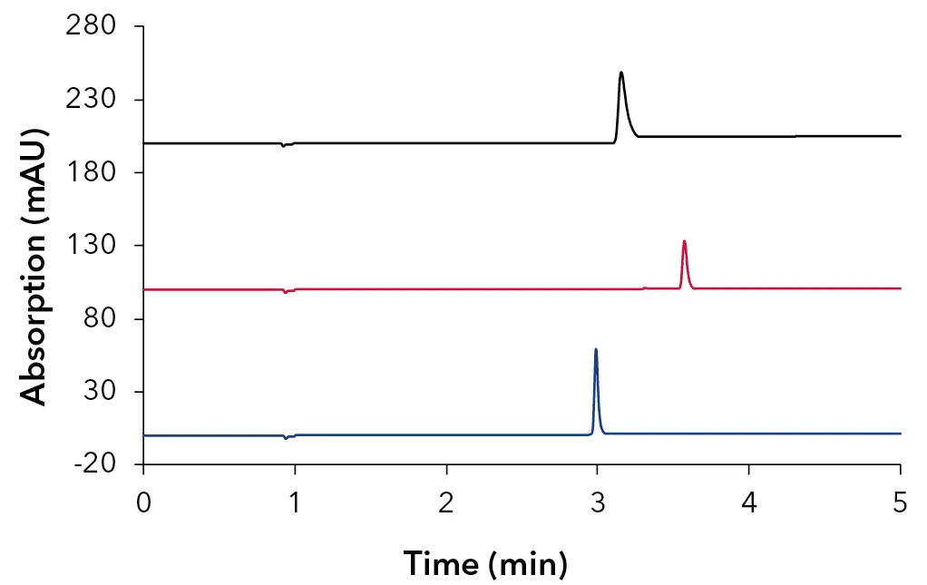
Fig. 1 Stacked overlay of oligonucleotide samples, 64mer – black, 25mer – red, 15mer – blue; 10 mM HAc + 10 mM TEA; 50 x 4.6 mm ID, 3 µm Sepapure oliGO column.
Sufficient retention as well as separation for all three oligonucleotide samples was achieved after optimizing the concentration of ion-pairing reagents and the gradient conditions. The ON were measured as single samples and not as a mixed sample because after a synthesis it is unlikely that you will have a mixture of ON with such a variety in length and number of base pairs.
Tab. 1 List of used oligonucleotide samples.
The optimized gradient was adapted to different column dimensions using the
KNAUER HPLC method converter and further optimized if necessary (Fig. 2). The following column dimensions were tested to investigate the On/Off retention mechanism of the used oligonucleotide samples in combination with the ion-pairing system consisting of HAc and TEA: 30 x 4.6 mm ID, 3 µm Sepapure oliGO column (Fig. 3/Tab. 2) and 10 x 4.6 mm, 3 µm Sepapure oliGO guard column (Fig. 4/Tab. 2).
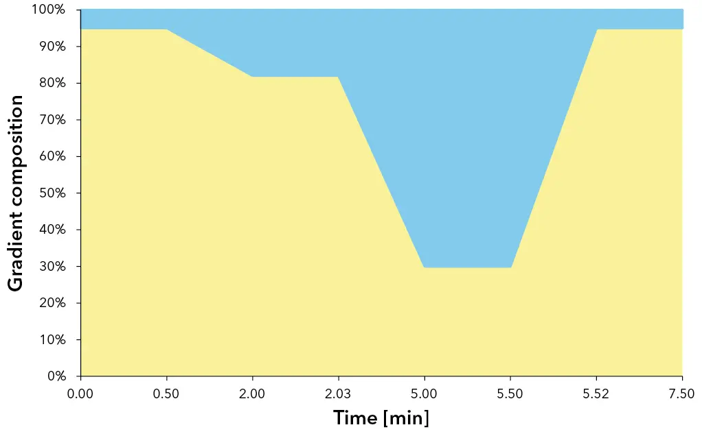
Fig. 2 Gradient composition and run time for 50 x 4.6 mm ID column.
Interestingly, the 64-mer ON elutes before the 25-mer. This is potentially caused by the 25-mer ON possessing a C12 linker-chain as a modification. This C12 chain contributes to interactions with the reversed phase column, resulting in a delayed elution of the 25-mer.
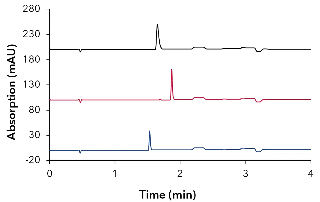
Fig. 3 Stacked overlay of oligonucleotide samples, 64mer – black, 25mer – red, 15mer – blue; 10 mM HAc + 10 mM TEA; 30 x 4.6 mm ID, 3 µm Sepapure oliGO column.
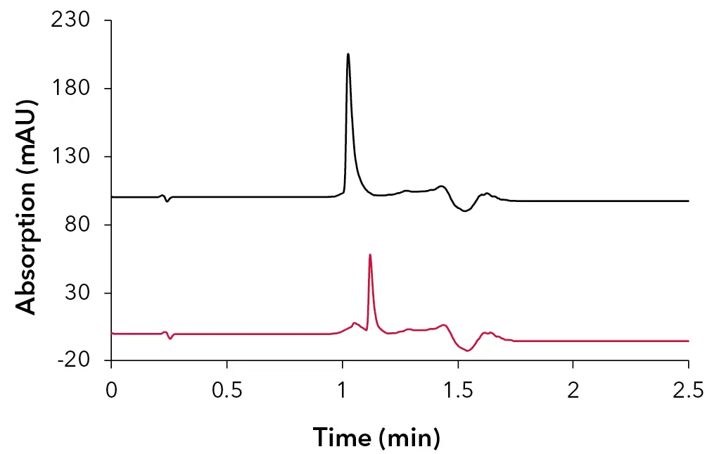
Fig. 4 Stacked overlay of oligonucleotide samples, 64mer – black, 25mer – red; 10 mM HAc + 10 mM TEA; 10 x 4.6 mm ID, 3 µm Sepapure oliGO column.
The downscaled gradient applied to the oliGO guard column worked well for the 64mer and the 25mer oligonucleotide sample. Unfortunately, the 15mer showed no retention on the 10 mm column. No further adjustments to the method were tested. Varying the ion-pairing reagent concentration or changing the gradient slope might be a possibility to achieve retention of the 15-mer oligonucleotide sample.
IP-RP system II – HFIP & TEA
In the next step, the developed methods were directly transferred to the second ion-pairing system, consisting of HFIP and TEA. For starting conditions, a concentration of 100 mM HFIP and 7 mM TEA (adjusted to pH 8) were chosen. Measurements were performed first on the 50 x 4.6 mm ID Sepapure oliGO column (Tab. 3). During the method optimization process, the concentration of TEA could be decreased to 4 mM while still maintaining a similar retention on the column. Fig. 5-7 show the separation of the three exemplary oligonucleotide samples using the second ion-pairing system.
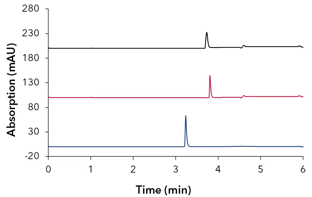
Fig. 5 Stacked overlay of oligonucleotide samples, 64mer – black, 25mer – red, 15mer – blue; 100 mM HFIP + 4 mM TEA; 50 x 4.6 mm ID, 3 µm Sepapure oliGO column.
The transfer of the gradient from one ion-pairing system to the other could be carried out without any adjustments for the 50 mm column.
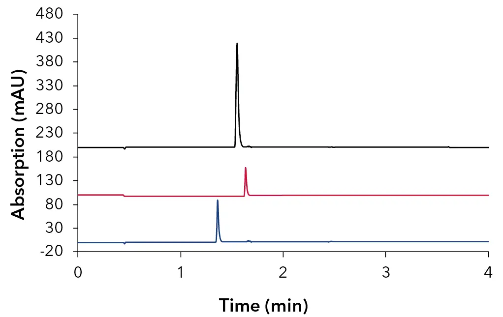
Fig. 6 Stacked overlay of oligonucleotide samples, 64mer – black, 25mer – red, 15mer – blue; 100 mM HFIP + 4 mM TEA; 30 x 4.6 mm ID, 3 µm Sepapure oliGO column, method parameters Tab. 4
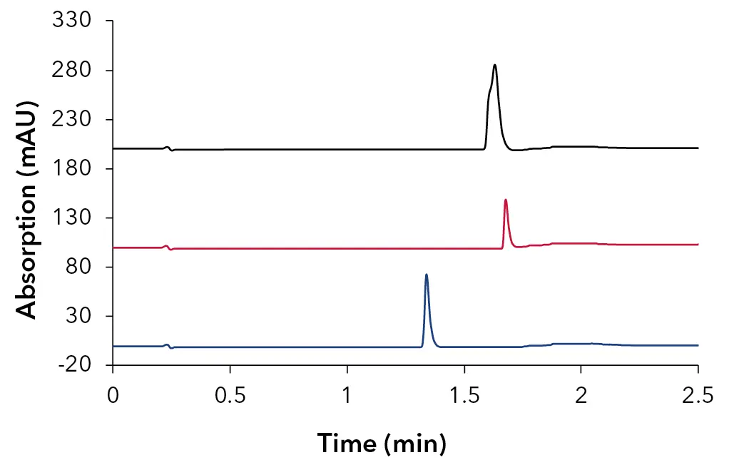
Fig. 7 Stacked overlay of oligonucleotide samples, 64mer – black, 25mer – red, 15mer – blue; 100 mM HFIP + 4 mM TEA; 10 x 4.6 mm ID, 3 µm Sepapure oliGO column, method parameters Tab. 4
Fazit
The experimental results demonstrate that HAc, in combination with TEA, constitutes a very suitable alternative to HFIP for IP-RP-HPLC with UV detection. While with the HFIP methods slightly shorter retention times were achieved HAc provided better peak symmetry, comparable resolution and overall chromatographic performance. The investigation into reduced column lengths confirmed the presence of a functional “On/Off” retention mechanism within the IP-RP-HPLC system. Both HFIP and HAc-based methods preserved sufficient retention and analyte resolution upon transfer to shorter columns. Furthermore, the used Sepapure oliGO column showed good overall symmetry even for higher molecular weight analytes (64mer). The ability to separate oligonucleotides in under two minutes is a good basis and shows the potential for high-throughput analysis.
Material und Methoden
Tab. 2 Method of oliGO columns for ion-pairing with HAc and TEA.
Tab. 3 Method of 50 mm oliGO column for ion-pairing with HFIP and TEA.
Tab. 4 Method for 30 mm/10 mm oliGO column for ion-pairing with HFIP and TEA.
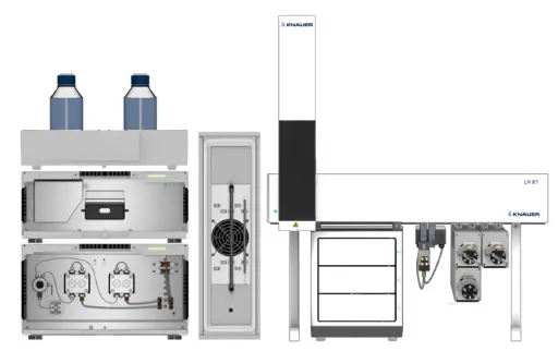
Tab. 5 System configuration.
References
[1] U.S. Department of Health and Human Services Food and Drug Administration Center for Drug Evaluation and Research (CDER); Clinical Pharmacology Considerations for the Development of Oligonucleotide Therapeutics Guidance for Industry; https://www.fda.gov/media/159414/download
[2] ECKSTEIN, F. Phosphorothioate Oligodeoxynucleotides: What Is Their Origin and What Is Unique about Them? Antisense and Nucleic Acid Drug Development 2000, 10 (2), 117–121. https://doi.org/10.1089/oli.1.2000.10.117.
[3] C. Michael Stein; C. Subasinghe; Kazuo Shinozuka; Cohen, J. S. Physicochemical Properties of Phosphorothioate Oligodeoxynucleotides. Nucleic Acids Research 1988, 16 (8), 3209–3221. https://doi.org/10.1093/nar/16.8.3209.
[4] DeVos, S. L.; Miller, T. M. Antisense Oligonucleotides: Treating Neurodegeneration at the Level of RNA. Neurotherapeutics 2013, 10 (3), 486–497. https://doi.org/10.1007/s13311-013-0194-5.
[5] Torgny Fornstedt, Martin Enmark; Separation of therapeutic oligonucleotides using ion-pair reversed-phase chromatography based on fundamental separation science; Journal of Chromatography Open, Volume 3, November 2023, 100079; https://doi.org/10.1016/j.jcoa.2023.100079
[6] Honorine LARDEUX, Selin BAGCI, Mimi GAO, Wiebke HOLKENJANS, Reinhard PELL, Davy GUILLARME; Understanding the fundamentals of the on-off retention mechanism of oligonucleotides and their application to high throughput analysis; Journal of Chromatography A Volume 1739, 4 January 2025, 465523; https://doi.org/10.1016/j.chroma.2024.465523
Anwendungsdetails