-

Connecting FPLC and structural analysis in SARS-CoV-2 research
Structural research requires pure specimen.
KNAUER supports your efforts with LC technology – for protein purification and more.

Structural research requires pure specimen.
KNAUER supports your efforts with LC technology – for protein purification and more.
Structural biology analyzes the structure of biomolecules and how alterations in their structures affect their function. Understanding the protein function on the molecular level gives valuable information for drug and vaccine development.
Several techniques are used to analyze the structure of proteins. Cryo-electron microscopy (cryo-EM) determines the high-resolution structure of biomolecules in solution by flash-freezing them and producing microscope images of individual molecules which are used to reconstruct the 3D structure of the molecule. Protein X-ray crystallography on the other hand uses x-ray diffraction of the crystallized biomolecules to obtain the 3D structure of a particular protein. Both techniques have in common that they depend on highly pure protein.

Numerous papers were published recently, reporting insights into the function of Covid19 proteins, shedding light on how the virus enters the cell and help to understand virus functioning. One of the important proteins is the surface spike glycoprotein of SARS-CoV-2. The spike protein binds to a receptor (ACE2, angiotensin converting enzyme II) on human cells causing the virus to enter cells and infect them. Several researchers reported structural data by cryo-EM or x-ray crystallography that will help to understand receptor recognition of SARS-CoV-2 (1-7). The important role of the spike protein for host cell infection makes it one of the key targets for medical intervention. The structural data will support the development of antiviral therapeutics and vaccines.
Another attractive drug target is the main protease (Mpro, 3CLpro) of SARS-CoV2. By inhibiting this protease, the virus replication can be prevented. Researchers reported the X-ray structures of the protease with and without an inhibitor. Based on their structure, they developed the lead compound into a potent inhibitor providing a valuable basis for the development of an anti-coronaviral drug (8).
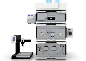
These proteins present major targets for vaccination and antiviral strategies underlining the importance of structural data of purified protein. One of the important aspects for structural analysis is the purification of proteins in solution. This is in almost all cases supported by fast liquid protein chromatography (FPLC). Most researchers use for their purification protocol a combination of affinity and size exclusion chromatography. Modern FPLC systems give researchers the freedom to automate their protein purification, increase reproducibility and therefor save time and resources. KNAUER offers a wide range of FPLC systems. Flexibility, modularity, and an easy scale up are the key benefits of our FPLC platform. Check out our preconfigured FPLC systems or get in touch with us to design a system fitting your needs.


for up to 50 mL/min
Article No.: SYS862113122
Show product
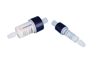
For affinity chromatography up to 50 ml/min
Article No.: A49031
Show product
Without first isolating and purifying proteins of interest it is difficult to precisely determine the three-dimensional (3D) structure of proteins. Protein purification can help researchers understand protein functionality.
Automate your purification and save time and resources. The Two-Step Purification System enables purifications over two steps, using two columns in one method.

FPLC is a form of liquid chromatography to purify large biomolecules like proteins. External factors like high temperature, high pressure, "extreme" pH, or solvents can disturb the protein structure and are therefore avoided in FPLC.
Prepacked FPLC columns and resins for all common protein purification applications: Size Exclusion Chromatography (SEC), Affinity Chromatography (AC) and Ion-Exchange Chromatography (IEX)
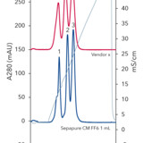
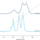
 Chromatogramm 280nm, B) SDS page (M - marker, F - filtrate, BT - breakthrough, W - wash, E - eluat, EEH - eluat treated with Endo H)")

Literature:
Wrapp D, Wang N, Corbett KS, et al. Cryo-EM structure of the 2019-nCoV spike in the prefusion conformation. Science. 2020;367(6483):1260–1263. doi:10.1126/science.abb2507
Shang J, Ye G, Shi K, et al. Structural basis of receptor recognition by SARS-CoV-2 [published online ahead of print, 2020 Mar 30]. Nature. 2020;10.1038/s41586-020-2179-y. doi:10.1038/s41586-020-2179-y
Yan R, Zhang Y, Li Y, Xia L, Guo Y, Zhou Q. Structural basis for the recognition of SARS-CoV-2 by full-length human ACE2. Science. 2020;367(6485):1444–1448. doi:10.1126/science.abb2762
Wang Q, Zhang Y, Wu L, et al. Structural and Functional Basis of SARS-CoV-2 Entry by Using Human ACE2 [published online ahead of print, 2020 Apr 7]. Cell. 2020;S0092-8674(20)30338-X. doi:10.1016/j.cell.2020.03.045
Walls AC, Park YJ, Tortorici MA, Wall A, McGuire AT, Veesler D. Structure, Function, and Antigenicity of the SARS-CoV-2 Spike Glycoprotein [published online ahead of print, 2020 Mar 6]. Cell. 2020;. doi:10.1016/j.cell.2020.02.058
Lan J, Ge J, Yu J, et al. Structure of the SARS-CoV-2 spike receptor-binding domain bound to the ACE2 receptor [published online ahead of print, 2020 Mar 30]. Nature. 2020;10.1038/s41586-020-2180-5. doi:10.1038/s41586-020-2180-5
Wang Q, Zhang Y, Wu L, et al. Structural and Functional Basis of SARS-CoV-2 Entry by Using Human ACE2 [published online ahead of print, 2020 Apr 7]. Cell. 2020;S0092-8674(20)30338-X. doi:10.1016/j.cell.2020.03.045
Anand K, Ziebuhr J, Wadhwani P, Mesters JR, Hilgenfeld R. Coronavirus main proteinase (3CLpro) structure: basis for design of anti-SARS drugs. Science. 2003;300(5626):1763–1767. doi:10.1126/science.1085658
Protein graphics used in header picture: Structure of the SARS-CoV-2 spike glycoprotein (closed state), by: Walls, A.C., Park, Y.J., Tortorici, M.A., Wall, A., Seattle Structural Genomics Center for Infectious Disease (SSGCID), McGuire, A.T., Veesler, D., doi: 10.2210/pdb6VXX/pdb
Molecule Images created using Mol* software (D. Sehnal, A.S. Rose, J. Kovca, S.K. Burley, S. Velankar (2018) Mol*: Towards a common library and tools for web molecular graphics MolVA/EuroVis Proceedings. doi:10.2312/molva.20181103) and RCSB PDB.